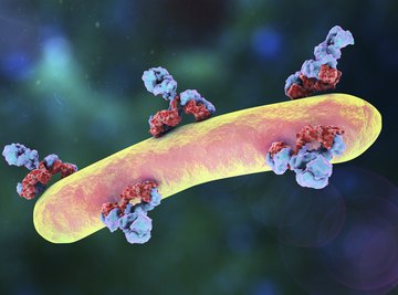

Immunoglobulins, also called antibodies, are glycoprotein molecules that make up an important part of the immune system, which is responsible for fighting off infectious disease and foreign "invasions" more generally. Often abbreviated as "Ig," antibodies are found in blood and other bodily fluids of humans and other vertebrate animals. They help identify and destroy foreign substances such as microbes (e.g., bacteria, protozoan parasites and viruses).
Immunoglobulins are classified into five categories: IgA, IgD, IgE, IgG and IgM. Only IgA, IgG and IgM are found in significant amounts in the human body, but all are important or potentially important contributors to the human immune response.
General Properties of Immunoglobulins
Immunoglobulins are produced by B-lymphocytes, which are a class of leukocytes (white blood cells). They are symmetrical Y-shaped molecules consisting of two longer heavy (H) chains and two shorter light (L) chains. Schematically, the "stem" of the Y includes the two L chains, which split apart about halfway from the bottom to the top of the immunoglobulin molecule and diverge at a roughly 90-degree angle. The two L chains run along the outsides of the "arms" of the Y, or the portions of the H chains above the split point. Thus, both the stem (two H chains) and both "arms" (one H chain, one L chain) consist of two parallel chains. The L chains come in two types, kappa and lambda. These chains all interact with each other via either disulfide (S-S) bonds or hydrogen bonds.
Immunoglobulins can also be separated into into constant (C) and variable (V) portions. The C portions direct activities in which all or most immunoglobulins participate, while the V areas bind to specific antigens (i.e., proteins that signal the presence of a particular bacteria, virus or other foreign molecule or entity). The "arms" of antibodies are formally called Fab regions, where "Fab" means "antigen-binding fragment"; the V portion of this includes only the first 110 amino acids of the Fab region, not the entire thing, as the portions of the Fab arms closest to the branch point of the Y are fairly constant between different antibodies and are considered part of the C region.
By way of analogy, consider a typical car key, which has a portion that is common to most keys regardless of the specific vehicle the key is designed to operate (e.g., the part you hold in your hand when using it) and a portion that is specific only to the vehicle in question. The handle portion can be likened to the C component of an antibody and the specialized portion to the V component.
Functions of the Constant and Variable Immunoglobulin Regions
The part of the C component below the branch of the Y, called the Fc region, may the thought of as the brains of the antibody operation. No matter what the V region is designed to do in a given type of antibody, the C region controls the executions of its functions. The C region of IgG and IgM is what activates the complement pathway, which are a set of nonspecific "first line of defense" immune responses involved in inflammation, phagocytosis (in which specialized cells physically engulf foreign bodies) and cell degradation. The C region of IgG binds to these phagocytes as well as to "natural killer" (NK) cells; the C region of IgE binds to mast cells, basophils and eosinophils.
As for particulars of the V region, this highly variable strip of the the immunoglobulin molecule is itself divided into hypervariable and framework regions. Diversity in the hypervariable reason, as your intuition probably suggests, is responsible for the amazing range of antigens that immunoglobulins are capable of recognizing, key-in-lock-style.
IgA
IgA accounts for about 15 percent of the antibodies in the human system, making it the second most common type of immunoglobulin. Only about 6 percent is found in the blood serum, however. In serum, it is found in its monomeric form – that is, as a single molecule in a Y shape as described above. In its secretory from, however, it exists as a dimer, or two of the Y monomers linked together. In fact, the dimeric form is more common, as IgA is seen in a wide variety of biological secretions, including milk, saliva, tears and mucus. It tends to be nonspecific in terms of the types of foreign presences it targets. Its presence on mucus membranes makes it an important gate-keeper in physically vulnerable locations, or the spots at which microbes might easily find ways deeper into the body.
IgA has a half-life of five days. The secretory form as a total of four sites at which to bind antigens, two per Y monomer. These are properly called epitope-binding sites, as the epitope is the specific part of any invader that triggers an immune reaction. Because it is found in mucous membranes that are exposed to high levels of digestive enzymes, IgA has a secretory component that prevents it from being degraded by these enzymes.
IgD
IgD makes up approximately 0.2 percent of the serum antibodies, or about 1 in 500. It is a monomer and has two epitope-binding sites.
IgD is found attached to the surface of B-lymphocytes as a B-cell receptor (also called sIg), where it is believed to control B-lymphocyte activation and suppression in response to signals from immumoglobulins that are circulating in blood plasma. IgD may be a factor in the active elimination of B-lymphocytes by generating self-reactive auto-antibodies. While it seems curious that antibodies would ever attack the cells that make them, sometimes this elimination may control an overzealous or misdirected immune response, or take B-cells out of the pool when they are damaged and no longer synthesizing helpful products.
In addition to its role as a de facto cell-surface receptor, IgD is found to a lesser extent in blood and lymphatic fluid. It is also thought in some people to react with certain haptens (antigenic subunits) on penicillin, which is likely why some people are allergic to this antibiotic; it may also react with ordinary, harmless blood proteins in the same way, thereby effecting an autoimmune response.
IgE
IgE is the rarest of the five classes of immunoglobulins, accounting for only about 0.002 percent of serum antibody, or about 1/50,000th of all circulating immunoglobulins. Nevertheless, it plays a vital role in the immune response.
Like IgD, IgE is a monomer and has two antigenic binding sites, one on each "arm." It has a short half-life of two days. It is bound to mast cells and basophils, which circulate in blood. As such, it is a mediator of allergic reactions. When an antigen binds to the Fab portion of an IgE molecule bound to a mast cell, this causes the mast cell to release histamine into the bloodstream. IgE also takes part in the lysis, or chemical degradation, of parasites of the protozoan variety (think amoebas and other unicellular or multicellular invaders). IgE is also made in response to the presence of helminths (parasitic worms) and certain arthropods.
At times, IgE also plays an indirect role in the immune response by galvanizing other immune components into action. IgE can protect mucosal surfaces by initiating inflammation. You may think that inflammation connotes something undesirable, since it tends to cause pain and swelling. But inflammation, among many its other immune benefits, enables IgG, which are proteins from the complement pathways, and white blood cells to enter tissues to confront invaders.
IgG
IgG is the dominant antibody in the human body, accounting for a whopping 85 percent of all immunoglobulins. Part of this is because of its long, albeit variable, half-life of seven to 23 days, depending on the IgG subclass in question.
Like three of the five types of immunoglobulin, IgG exists as a monomer. It is found chiefly in the blood and lymph. It has the unique ability to cross placenta in pregnant women, allowing it to protect the unborn fetus and newborn baby. Its main activities include enhancing phagocytosis in macrophages (specialized "eater" cells) and neutrophils (another type of white blood cell); neutralizing toxins; and inactivating viruses and killing bacteria. This gives IgG a wide palette of functions, fitting for an antibody that is so prevalent in the system. It is usually the second antibody on the scene when an invader is present, following closely behind IgM. Its presence is vastly increased in the body's anamnestic response. "Anamnestic" translates to "not forgetting," and IgM responds to an invader that it has encountered before with an immediate spike in its numbers. Finally, the Fc portion of IgG can bind to NK cells to set in motion a process called antibody-dependent cell-mediated cytotoxicity, or ADCC, which can kill or limit the effects of invading microbes.
IgM
IgM is the colossus of immunoglobulins. It exists as a pentameter, or a group of five bound IgM monomers. IgM has a short half life (about five days) and makes up approximately 13 to 15 percent of serum antibodies. Importantly, it is also the first line of defense among its four antibody siblings, being the first immunoglobulin made during a typical immunological response.
Because IgM is a pentamer, it has 10 epitope-binding sites, making it a fierce adversary. Its five Fc portions, like those of most other immunoglobulins, can activate the complement-protein pathway, and as a "first responder" is the most efficient type of antibody in this regard. IgM agglutinates invading material, compelling individual pieces to stick together for easier clearing from the body. It also promotes lysis and phagocytosis of micro-organisms, with a particular affinity for ousting bacteria.
Monomeric forms of IgM do exist and are found chiefly on the surface of B-lymphocytes as receptors or sIg (as with IgD). Interestingly, the body has already produced adult levels of IgM by the age of nine months.
A Note on Antibody Diversity
Thanks to the very high variability of the hypervariable portion of the Fab component of each of the five immunoglobulins, an astronomical number of unique antibodies can be created across the five formal classes. This is augmented by the fact that the L and H chains also come in a number of isotypes, or chains that are superficially the same in arrangement but contain different amino acids. In fact, there are 45 different "kappa" L chain genes, 34 "lambda" L chain genes and 90 H chain genes for a total of 177, in turn yielding over three million unique combinations of genes.
This makes sense from the standpoint of evolution and survival. Not only must the immune system be prepared to confront invaders it already "knows" about, but it also must be prepared to create an optimal response to invaders it has never seen or, for that matter, that are brand-new in nature, such as influenza viruses that have evolved themselves through mutations. The host-invader interaction over time and across microbial and vertebrate species is really no more than an ongoing, interminable "arms race."
References
Resources
About the Author
Kevin Beck holds a bachelor's degree in physics with minors in math and chemistry from the University of Vermont. Formerly with ScienceBlogs.com and the editor of "Run Strong," he has written for Runner's World, Men's Fitness, Competitor, and a variety of other publications. More about Kevin and links to his professional work can be found at www.kemibe.com.