What does fact checked mean?
At Healthfully, we strive to deliver objective content that is accurate and up-to-date. Our team periodically reviews articles in order to ensure content quality. The sources cited below consist of evidence from peer-reviewed journals, prominent medical organizations, academic associations, and government data.
The information contained on this site is for informational purposes only, and should not be used as a substitute for the advice of a professional health care provider. Please check with the appropriate physician regarding health questions and concerns. Although we strive to deliver accurate and up-to-date information, no guarantee to that effect is made.
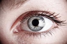
What Causes Calcium Deposits in the Eye?
Calcium deposits can form in three different places in the eye--the cornea, conjunctiva and retina. The cornea is the clear tissue on your eyeball that covers your iris and pupil. The conjunctiva is the clear membrane that surrounds the white part of your eyeball and the inside of your eyelids. The retina is the nerve tissue in the back of your eyeball that detects light as your eye collects it.
Cornea
The cornea will exhibit calcium deposits in a condition known as band keratopathy. According to Wills Eye Hospital, this type of calcium deposit is the result of hypercalcemia, gout or renal failure. Each of these three conditions results in excess calcium levels in the body. The excess calcium is deposited in the cornea as it is transported there in the blood vessels. Chronic inflammation of the eyeball and exposure to toxic vapors such as mercury can also cause band keratopathy to develop. The treatment of band keratopathy involves the treatment of the underlying condition. It can also involve the instillation of artificial tear drops. In the most severe cases, a chelation agent is used. Chelation agents are applied to an area of the body to remove minerals such as calcium from the affected tissue.
- The cornea will exhibit calcium deposits in a condition known as band keratopathy.
- According to Wills Eye Hospital, this type of calcium deposit is the result of hypercalcemia, gout or renal failure.
Conjunctiva
High Cholesterol Effects on Eyes
Learn More
The conjunctiva will exhibit calcium deposits in the form of concretions. Concretion.org explains that a concretion is a small, hard, yellow or white deposit. This deposit forms in the conjunctiva that lines the inside of your eyelids. Concretion.org states that concretions are most common if you have a condition that causes your eyes to be inflamed for a long period of time. The cells in your conjunctiva begin to degenerate. As this happens, other materials fill the space in what is known as a cyst. As the cyst hardens, it forms a small piece of calcium. The treatment of large concretions is to have your optometrist of ophthalmologist remove them. This requires only a topical anesthetic.
- The conjunctiva will exhibit calcium deposits in the form of concretions.
- This deposit forms in the conjunctiva that lines the inside of your eyelids.
Retina
The retina will exhibit calcium deposits in the form of calcific plaques in the retinal arteries 3. These plaques are visible to your optometrist or ophthalmologist as they view the inside of your eyeball. Wills Eye Hospital explains that these calcific plaques break off of a damaged heart valve 3. The plaque is then transported through your arteries until it reaches an artery that is too small for it to pass through. It becomes lodged in the wall of that artery. The calcific plaque can become lodged in a small artery anywhere in the body. These plaques are most readily visible in the retina because the small arteries can be viewed during a routine eye examination. They can cause the artery to become clogged. If the affected artery is large enough, it can cause vision loss. The treatment for a calcific plaque depends on its size and location. Your optometrist or ophthalmologist will direct you toward the appropriate treatment depending on your unique needs.
- The retina will exhibit calcium deposits in the form of calcific plaques in the retinal arteries 3.
- The calcific plaque can become lodged in a small artery anywhere in the body.
Related Articles

High Cholesterol Effects on Eyes
Learn More

What Eye Problems are Typical With Crohn's Disease?
Learn More
Calcium and the Optic Nerve
Learn More

What Causes Eye Floaters in Vision?
Learn More

What Causes a Kaleidescope Halo in the Eye?
Learn More

Types of Growths on the Eye
Learn More

List of Medications to Avoid With Glaucoma
Learn More

How to Heal Floaters With Enzymes
Learn More
How to Get Rid of Swelling on the Inner Corner of My Eye
Learn More

Foods to Help Prevent Cataracts
Learn More
References
- "Wills Eye Hospital: Cornea": Rapuano, Christopher J., M.D., Heng, Wee-Jin, M.D.: 2003
- "Wills Eye Hospital: Retina": Ho, Allen C., M.D., Brown, Gary C, M.D., McNamara, J. Arch, M.D., Recchia, Franco, M., M.D., Regillo, Carl D., M.D., Vander, James F., M.D.: 2003
- Heart: Calcific Retinal Embolism
- Merolla G, Singh S, Paladini P, Porcellini G. Calcific tendinitis of the rotator cuff: state of the art in diagnosis and treatment. J Orthop Traumatol. 2016;17(1):7–14. doi:10.1007/s10195-015-0367-6
- DE Carli A, Pulcinelli F, Rose GD, Pitino D, Ferretti A. Calcific tendinitis of the shoulder. Joints. 2014;2(3):130–136. doi:10.11138/jts/2014.2.3.130
- Southern California Orthopedic Institute. Calcium deposits in the shoulder.
- Suzuki K, Potts A, Anakwenze O, Singh A. Calcific tendinitis of the rotator cuff: management options. J Am Acad Orthop Surg. 2014 Nov;22(11):707-17. doi:10.5435/JAAOS-22-11-707
Writer Bio
Travis Sharpe began writing in 2009. His articles appear on eHow and Answerbag, where he specializes in eye health, general health and prescription medications. Sharpe is a Fellow of the American Academy of Optometry (AAO) and graduated summa cum laude from his alma mater. He has a Doctor of Optometry from the Southern College of Optometry.