
Taking a peek into a microscope can take you to a different world. The ways microscopes zoom in on objects at a small scale are similar to how glasses and magnifying glasses can let you see better.
Compound microscopes in particular work using an arrangement of lenses for refracting light to zoom in on cells and other specimens to take you into a micro-sized world. A microscope is called a compound microscope when it consists of more than one set of lenses.
Compound microscopes, also known as optical or light microscopes, work by making an image appear much larger through two systems of lenses. The first is the ocular, or eyepiece lens, that you look into when using the microscope that typically magnifies at a range between five times and 30 times. The second is the objective lens system that zooms in using magnitudes from four times up to 100 times, and compound microscopes usually have three, four or five of these.
Lenses in a Compound Microscope
The objective lens system use a small focal distance, the distance between the lens and the specimen or object being examined. The real image of the specimen is projected through the objective lens to create an intermediate image from the light incident on the lens that is projected onto the objective conjugate image plane or the primary image plane.
Changing objective lens magnification changes how this image is scaled up in this projection. The optical tube length refers to the distance from the back focal plane of the objective to the primary image plane within the microscope body. The primary image plane is usually either within the microscope body itself or within the eyepiece.
The real image is then projected onto the eye of the person using the microscope. The ocular lens does this as a simple magnifying lens. This system from objective to ocular shows how the two lens systems work one after the other.
The compound lens system lets scientists and other researchers create and study images at a much higher magnification that they could otherwise only achieve with one microscope. If you were to try to use a microscope with a single lens to achieve these magnifications, you would have to place the lens very close to your eye or use a very wide lens.
Dissecting Microscope Parts and Functions
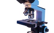
Dissecting microscope parts and functions can show you how they all work together when studying specimens. You can roughly divide sections of the microscope into the head or body, base and arm with the head at the top, the base at the bottom and the arm in between.
The head has an eyepiece and eyepiece tube that holds the eyepiece in place. The eyepiece can be either monocular or binocular, the latter of which can use a diopter adjustment ring to make the image more consistent.
The arm of the microscope contains the objectives that you can choose and place for different levels of magnification. Most microscopes use 4x, 10x, 40x and 100x lenses that work as coaxial knobs controlling how many times the lens magnifies the image. This means they're built on the same axis as the knob that's used for fine focus, as the word "coaxial" would imply. The objective lens in microscope function
At the bottom is the base that supports the stage and the light source that projects through an aperture and lets the image project through the rest of the microscope. Higher magnifications usually use mechanical stages that let you use two different knobs to move both left and right and forward and backward.
The rack stop lets you control the distance between the objective lens and the slide for an even closer look at the specimen.
Adjusting the light coming from the base is important. Condensers receive the incoming light and focus it onto the specimen. The diaphragm lets you choose how much light reaches the specimen. The lenses in a compound microscope use this light in creating the image for the user. Some microscopes use mirrors to reflect light back onto the specimen instead of a light source.
Ancient History of Microscope Lenses
Humans have studied how glass bends light for centuries. Ancient roman mathematician Claudius Ptolemy used mathematics to explain the precise angle of refraction about how the image of a stick refracted when placed into water. He would use this to determine the refraction constant or index of refraction for water.
You can use the index of refraction to determine how much the speed of light changes when passed into another medium. For a particular medium, use the equation for index of refraction
for index of refraction n, speed of light in a vacuum c (3.8 x 10⁸ m/s) and speed of light in the medium v.
The equations shows how light slows down when entering media such as glass, water, ice or any other medium whether it's solid, liquid or gas. Ptolemy's work would prove essential to microscopy as well as optics and other areas of physics.
You can also use Snell's law to measure the angle at which a beam of light refracts when it enters a medium, much the same way Ptolemy deduced. Snell's law is
for θ1 as the angle between the line of the beam of light and the line of the edge of the medium before light enters the medium and θ2 as the angle after light has entered. n1 and n2 are the indices of refraction for the medium light was previously in and the medium light enters.
As more research was done, scholars began taking advantage of the properties of glass around the first century AD. By that time, Romans had invented glass and began testing it for its uses in magnifying what can be seen through it.
They started experimenting with different shapes and sizes of glasses to figure out the best way to magnify something by looking through it including how it could direct the sun's rays to light objects on fire. They called these lenses "magnifiers" or "burning glasses."
The First Microscopes
Near the end of the 13th century, people started creating glasses using lenses. In 1590, two Dutch men, Zaccharias Janssen and his father Hans, performed experiments using the lenses. They discovered that placing the lenses one on top of the other in a tube could enlarge an image at much greater magnification than a single lens could achieve, and Zaccharias soon invented the microscope. This similarity to the objective lens system of microscopes shows how far back the idea of using lenses as a system goes.
The Janssen microscope used a brass tripod about two and a half feet long. Janssen fashioned the primary brass tube that the microscope used at around an inch or half of an inch in radius. The brass tube had discs at the base as well as at each end.
Other microscope designs began to arise by scientists and engineers. Some of them used a system of a large tube that housed two other tubes that slid into them. These handmade tubes would magnify objects and serve as the basis for the design of modern microscopes.
These microscopes weren't usable for scientists just yet, though. They would magnify images about nine times while leaving the images they created difficult to see. Years later, by 1609, astronomer Galileo Galilei was studying the physics of light and how it would interact with matter in ways that would prove beneficial to the microscope and telescope. He also added a device to focus the image to his own microscope.
Dutch scientist Antonie Philips van Leeuwenhoek used a single-lens microscope in 1676 when he would use small glass spheres to become the first human to observe bacteria directly, becoming known as "the father of microbiology."
When he looked at a drop of water through the lens of the sphere, he saw the bacteria floating around in the water. He would go on to make discoveries in plant anatomy, discover blood cells and make hundreds of microscopes with new ways of magnifying. One such microscope was able to use magnification at 275 times using a single lens with a double-convex magnifier system.
Advances in Microscope Technology
The coming centuries brought more improvements to microscope technology. The 18th and 19th centuries saw refinements to microscope designs to optimize efficiency and effectiveness, such as making the microscopes themselves more stable and smaller. Different lens systems and power of lenses themselves addressed the issues of the blurriness or lack of clarity in images that microscopes produced.
The advances in the optics of science brought a greater understanding of how images are reflected onto different planes that lenses could create. This let the creators of microscopes create more precise images during these advances.
In the 1890s, then-German graduate student August Köhler published his work on Köhler illumination that would distribute light to reduce optical glare, focus light on the subject of the microscope and use more precise methods of controlling the light in general. These technologies relied on the index of refraction, size of the aperture contrast between the specimen and the light of the microscope alongside more control the components such as the diaphragm and eyepiece.
Lenses of Microscopes Today
Lenses today vary from ones that focus on specific colors to lenses that apply to certain refractive indices. Objective lens systems use these lenses to correct for chromatic aberration, color disparities when different colors of light differ slightly in the angle at which they refraction. This occurs due to the differences in wavelength of different colors of light. You can figure out which lens is appropriate for what you want to study.
Achromatic lenses are used to make refractive indices of two different wavelengths of light the same. They're generally priced at an affordable rate and, as such, are widely used. Semi-apochromatic lenses, or fluorite lenses, change the refractive indices of three wavelengths of light to make them the same. These are used in studying fluorescence.
Apochromatic lenses, on the other hand, use a large aperture for letting light through and achieve a higher resolution. They're used for detailed observations, but they're usually more expensive. Plan lenses address the effect of field curvature aberration, the loss in focus when a curved lens creates the sharpest focus of an image away from the plane it's meant to project the image upon.
Immersion lenses increase the aperture size using a liquid that fills the space between the objective lens and the specimen, which also increases the resolution of the image.
With advances in technology of lenses and microscopes, scientists and other researchers determine the precise causes of disease and specific cellular functions that governed biological processes. Microbiology showed a whole world of organisms beyond the naked eye that would lead to more theorizing and testing of what it meant to be an organism and what the nature of life was like.
References
- Microscope: Compound Microscope Parts
- Microbus: Microscope Parts & Specifications
- BBC: Antonie van Leeuwenhoek (1632 - 1723)
- New York Microscope Company: What is a Compound Microscope?
- Microscope World: Microscope History - Who Invented the Microscope?
- Sciencedirect: Microscopy
- Hyperphysics: Refraction of Light
- Vision Engineering: History of the Microscope
- MicroscopeMaster: History of the Microscope
- Olympus: Köhler Illumination
- Keyence: Microscope Optical Systems
About the Author
S. Hussain Ather is a Master's student in Science Communications the University of California, Santa Cruz. After studying physics and philosophy as an undergraduate at Indiana University-Bloomington, he worked as a scientist at the National Institutes of Health for two years. He primarily performs research in and write about neuroscience and philosophy, however, his interests span ethics, policy, and other areas relevant to science.