What does fact checked mean?
At Healthfully, we strive to deliver objective content that is accurate and up-to-date. Our team periodically reviews articles in order to ensure content quality. The sources cited below consist of evidence from peer-reviewed journals, prominent medical organizations, academic associations, and government data.
The information contained on this site is for informational purposes only, and should not be used as a substitute for the advice of a professional health care provider. Please check with the appropriate physician regarding health questions and concerns. Although we strive to deliver accurate and up-to-date information, no guarantee to that effect is made.
Dark Spots on the Brain
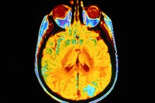
A dark spot can appear on an X-ray or scan for any number of reasons. Brain lesions usually are discovered accidentally when you're being diagnosed for an unrelated symptom, according to MayoClinic.com 1. Dark spots that indicate brain lesions usually are discovered after undergoing a magnetic resonance imaging test, or MRI, or a CT scan, otherwise called a computerized tomography scan 1.
Causes
A dark spot on the brain can be a leftover stain from an old injury or resolved medical condition and pose no threat. On the other hand, a lesion that appears on a test as a dark stain can indicate a number of serious conditions varying from a tumor to an aneurysm or congenital brain abnormality. Further tests and examinations may be required to diagnose the spot, or your doctor may choose to monitor the lesion to watch for changes.
Features
Life Expectancy for Spinal & Brain Cancer
Learn More
A brain tumor that originates in the brain is called a primary brain tumor. Cancer that has spread to the brain from another part of the body is called metastatic brain tumors, according to the National Cancer Institute 2. Other neurological symptoms such as headaches or vision loss may prompt further tests to look for dark spots. A dye may be injected in the bloodstream to highlight the potential brain tumor.
- A brain tumor that originates in the brain is called a primary brain tumor.
- A dye may be injected in the bloodstream to highlight the potential brain tumor.
Effects
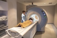
An MRI is the most commonly used technique when diagnosing brain lesions 1. Advances in MRI technology allow doctors to see with greater detail how much damage is associated with the dark spot and what the underlying disease may be that's causing the abnormality. An MRI is performed in a large tube-shaped machine that you enter while resting on a movable table. Loud banging noises occur throughout the procedure that can last up to an hour while the scans are taken.
- An MRI is the most commonly used technique when diagnosing brain lesions 1.
- Loud banging noises occur throughout the procedure that can last up to an hour while the scans are taken.
Considerations
What Is Stage 4 Brain Cancer?
Learn More
While symptoms of a tumor or lesion on the brain may prompt your doctor to order scans of your brain, the only definitive way to identify a tumor is through a biopsy, according to the American Brain Tumor Association. X-rays and scans can indicate the presence of a dark spot and help to determine the general skull condition surrounding the tumor, guiding surgeons in their probes. Images can help to provide preliminary diagnoses and direct further tests.
- While symptoms of a tumor or lesion on the brain may prompt your doctor to order scans of your brain, the only definitive way to identify a tumor is through a biopsy, according to the American Brain Tumor Association.
- X-rays and scans can indicate the presence of a dark spot and help to determine the general skull condition surrounding the tumor, guiding surgeons in their probes.
Identification
To concretely identify the dark spot as a malignant brain tumor, other tests usually are provided, notes the National Cancer Institute 2. A biopsy is performed by making a small incision in the skull and taking out a piece of the spot to test it under a microscope and in the laboratory. A spinal tap that removes fluid from the top of the spine that leads to the brain also may help identify if cancer is present in the tumor.
Related Articles
Life Expectancy for Spinal & Brain Cancer
Learn More
What Is Stage 4 Brain Cancer?
Learn More
What Causes Calcium Deposits on the Brain?
Learn More

Why Is Creatine Level Important for a Brain MRI?
Learn More
Life Expectancy for Spinal & Brain Cancer
Learn More

How to Tell If Plastic Is BPA Free
Learn More

What Does a Pinched Nerve MRI Look Like?
Learn More

Age Spots and Moles
Learn More
Protein, Ammonia & Brain Damage
Learn More
Side Effects of MRI With Contrast
Learn More
References
- MayoClinic.com: Brain Lesions
- National Cancer Institute: Brain Tumors
- National Institute of Neurological Disorders and Stroke. Brain and Spinal Cord Tumors: Hope Through Research. Updated December 31, 2019.
- American Cancer Society. Tests for Brain and Spinal Cord Tumors in Adults. Updated November 6, 2017.
- American Cancer Society. Types of Brain and Spinal Cord Tumors in Adults. Updated January 21, 2016.
- American Cancer Society. What Are Pituitary Tumors? Updated November 2, 2017.
- Le Rhun E, Taillibert S, Chamberlain MC. Carcinomatous meningitis: Leptomeningeal metastases in solid tumors. Surg Neurol Int. 2013;4(Suppl 4):S265–S288. doi:10.4103/2152-7806.111304
- American Society of Clinical Oncology. Brain Tumor: Grades and Prognostic Factors. Updated March 2019.
- Patel K, Clifford DB. Bacterial brain abscess. Neurohospitalist. 2014;4(4):196–204. doi:10.1177/1941874414540684
Writer Bio
Linda Ray is an award-winning journalist with more than 20 years reporting experience. She's covered business for newspapers and magazines, including the "Greenville News," "Success Magazine" and "American City Business Journals." Ray holds a journalism degree and teaches writing, career development and an FDIC course called "Money Smart."