
Gel electrophoresis is a method used in laboratories to measure and sort strands of DNA. It is necessary because DNA under normal conditions is too small to manipulate, even when viewed using most microscopes. The gel electrophoresis lab uses a relatively straightforward procedure, and the same basic technique can be used to separate individual proteins, as well.
The Gel Matrix
To begin the gel electrophoresis procedure, you first must create the gel. Typically, gels are made in thin sheets using a substance called agarose. Powdered agarose is placed in a flask, followed by a salt water solution called a buffer. This mixture of agarose and buffer is heated until the two substances melt together, then poured into a forming mold. A device called a comb is then placed at one end of the mold before the gel cools. When the gel is cooled, the comb is removed, leaving little slots which will be used to hold DNA samples.
A special characteristic of the cooled agarose mixture (called a gel matrix) stems from the fact that it is created with salt water. When electrified, the matrix will become conductive, allowing electricity to flow along its length. Another special property of the gel matrix is the presence of regular, microscopic holes. These holes will allow strands of DNA to travel through the gel matrix and facilitate the sorting process.
The Electrophoresis Chamber
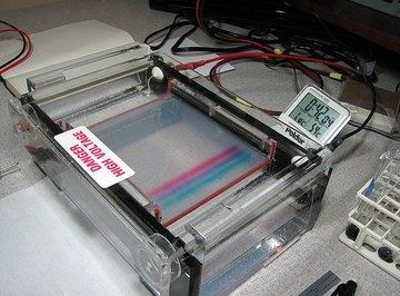
Your next step is to create an electrophoresis chamber. This is a small rectangular box, wired with a positive and negative electrical connection at either end. Chambers are typically shallow, small enough to fit on a tabletop, and built from clear materials like Plexiglas.
Salt water solution is poured into the bottom of the electrophoresis chamber, and the gel matrix is submerged slightly within this solution. The salt water serves two purposes: aiding the flow of electricity and keeping the gel matrix moist. Since DNA is propelled by a negative charge, place your matrix so your samples will be located next to your negative electrical connection.
Preparing the DNA
DNA samples are then prepared. Since DNA in solution is all but impossible to see, a coloring agent called a loading buffer is added to each individual sample. This agent also thickens the DNA solution, making it less runny and more workable. Using a pipette, transfer a sample of DNA solution into each alternating slot in the gel matrix. In the empty slot between each sample, place some solution of DNA whose length you already know (called DNA standard) for experiment control and comparison.
Turn On the Power
Now, turn on your electrophoresis chamber. Under negative power, your DNA samples will be forced across the length of the chamber. Small strands of DNA will move more quickly through the gel matrix, and in a short amount of time they will separate themselves from longer, slower strands. The dye in the coloring agent lets you follow the track of the DNA. You will not be able to see individual strands of DNA, but strands of the same length will clump together.
Final Steps
When the DNA is sorted out, the matrix is removed from the electrophoresis chamber. The DNA is then stained to allow for easier measurement and examination.
References
About the Author
M. Gideon Hoyle is a writer living outside of Houston. Previously, he produced brochures and a wide variety of other materials for a nonprofit educational foundation. He now specializes in topics related to health, exercise and nutrition, publishing for various websites.