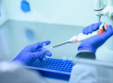

Gel electrophoresis is a technique that allows DNA to be analyzed at the level of its constituent molecules. In this DNA visualization method, samples are placed on an agarose gel medium and an electric field is applied to the gel. This causes fragments of DNA to migrate through the gel at different rates in accordance with their electrochemical properties.
Ethidium Bromide
For this visualization technique, ethidium bromide is mixed with agarose powder, EDTA buffer and water to form the gel matrix before electrophoresis. As a result, the ethidium bromide molecules become uniformly dispersed throughout the matrix. Once the gel's wells have been filled with their respective DNA samples and tracking dyes, voltage is applied to slowly draw the large, polar compounds across the matrix.
During this movement, the bases of the DNA molecules temporarily bind to the particles thanks to the ethidium bromide charge, dragging them along. By the time gel electrophoresis is complete, each DNA molecule has picked up at significant amount of ethidium bromide.
In the presence of ultraviolet light, ethidium bromide exhibits fluorescence. Technicians shine a specially-calibrated UV light across the gel while a machine captures the picture of the glowing fragments.
Methylene Blue
If a UV transilluminator is not available or practical, technicians can render DNA visible under normal condition by soaking the finished agarose gel, with electrophoresized DNA inside, in a solution of methylene blue overnight.
A chloride salt with a significantly hydrophobic anion, methylene blue molecules penetrate the entire gel matrix. However, the hydrogen bonding throughout DNA causes the stain molecules to accumulate. This increased DNA stain density yields a deeper shade of blue, visible to the naked eye.
Tracking Dyes
Beyond the relative size of the DNA bands, technicians can measure the absolute size (in base-pairs) of each fragment using chemicals called tracking dyes. Visible without the addition of methylene blue or ethidium bromide, tracking dyes such as bromophenol blue and xylene cyanol move across the aragose gel matrices during electrophoresis at the same speed as DNA fragments consisting of 300 nucleotides and 4,000 nucleotides, respectively. In electrophoresis, more massive DNA fragments travel across the gel matrix at a slower speed than smaller fragments. Therefore, while tracking dyes do not directly influence the visibility of DNA fragments, comparing the position of a DNA fragment in the gel to the position of these dyes allow technicians to "see" the approximate number of nucleotides the DNA fragment contains.
References
About the Author
A Chicago-based copywriter, Andy Pasquesi has extensive experience writing for automotive (BMW, MINI Cooper, Harley-Davidson), financial services (Ivy Funds, William Blair, T. Rowe Price, CME Group), healthcare (Abbott) and consumer goods (Sony, Motorola, Knoll) clients. He holds a Bachelor of Arts in English from Harvard University but does not care for the Oxford comma.