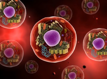
Cells are the fundamental, irreducible elements of life on Earth. Some living things, such as bacteria, consist of only a single cell; animals such as yourself include trillions. Cells are themselves microscopic, yet most of them contain a staggering array of even smaller components that all contribute to the basic mission of keeping the cell – and by extension, the parent organism – alive. Animal cells are, generally speaking, part of more complex life forms than are bacterial or plant cells; accordingly, animal cells are more complicated and elaborate than their counterparts in the microbial and botanical worlds.
Perhaps the easiest way to think of an animal cell is as a fulfillment center or large, busy warehouse. An important consideration to keep closely in mind, one that often describes the world in general but is exquisitely applicable to biology in particular, is "form fits function." That is, the reason why the parts of an animal cell, as well as the cell as whole, are structured the way they are is very closely related to the jobs these parts – called "organelles" – are tasked with carrying out.
Basic Overview of Cells
Living things can be divided into prokaryotic organisms, which are single-celled and include:
- plants
- animals
- fungi
The cells of eukaryotes include a membrane around the genetic material, creating a nucleus; prokaryotes have no such membrane. Also, the cytoplasm of prokaryotes contains no organelles, which eukaryotic cells boast in abundance.
The Animal Cell Membrane
The cell membrane, also called the plasma membrane, forms the outer boundary of animal cells. (Plant cells have cell walls directly outside the cell membrane for added protection and firmness.) The membrane is more than a simple physical barrier or a warehouse for organelles and DNA; instead, it is dynamic, with highly selective channels that carefully regulate the entry and exit of molecules to and from the cell.
The cell membrane consists of a phospholipid bilayer, or lipid bilayer. This bilayer consists of, in essence, two different "sheets" of phospholipid molecules, with the lipid parts of the molecules in different layers touching and the phosphate parts pointing in opposite directions. To understand why this occurs, consider the electrochemical properties of lipids and phosphates separately. Phosphates are polar molecules, meaning that their electrochemical charges are distributed unevenly across the molecule. Water (H2O) is also polar, and polar substances tend to commingle, so phosphates are among the substances labeled hydrophilic (i.e., attracted to water).
The lipid portion of a phospholipid contains two fatty acids, which are long chains of hydrocarbons with specific types of bonds that leave the whole molecule without a charge gradient. In fact, lipids are by definition nonpolar. Because they react opposite to the way that polar molecules do in the presence of water, they are called hydrophobic. You might therefore think of a whole phospholipid molecule as "squid-like," with the phosphate part serving as the head and body and the lipid as a pair of tentacles. Further, imagine two large "sheets" of squids, gathered with their tentacles mingling and their heads pointed in opposite directions.
Cell membranes allow certain substances to come and go. This occurs in a number of ways, including diffusion, facilitated diffusion, osmosis and active transport. Some organelles, such as mitochondria, have their own internal membranes consisting of the same materials as the plasma membrane itself.
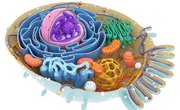
The Nucleus
The nucleus is, in effect, the control and command center of the animal cell. It contains the DNA, which in most animals is arranged in separate chromosomes (you have 23 pairs of these) that are divided into small portions called genes. Genes are simply lengths of DNA that contain the code for a particular protein product, which the DNA delivers to the cell's protein-assembly machinery through the molecule RNA (ribonucleic acid).
The nucleus includes different portions. On microscopic examination, a dark spot called the nucleolus appears in the middle of the nucleus; the nucleolus is involved in the manufacture of ribosomes. The nucleus is surrounded by a nuclear membrane, a double later analogous to the cell membrane. This lining, also called the nuclear envelope, has filamentous proteins attached to the inside layer that extend inward and help keep DNA organized and in place.
During cell reproduction and division, the cleavage of the nucleus itself into two daughter nuclei is called cytokinesis. Having the nucleus separate from the rest of the cell is useful in keeping the DNA isolated from other cell activities, minimizing the chances that it might be damaged. This also allows for exquisite control of the immediate cellular environment, which can be distinct from the cytoplasm of the cell at large.
Ribosomes
These organelles, which are also found in non-animal cells, are responsible for protein synthesis, which occurs in the cytoplasm. Protein synthesis is set in motion when DNA in the nucleus undergoes a process called transcription, which is the making of RNA with a chemical code corresponding to the exact strip of DNA from which it is made (messenger RNA or mRNA). DNA and RNA both consist of monomers (single repeating units) of nucleotides, which contain a sugar, a phosphate group and a portion called a nitrogenous base. DNA includes four different such bases (adenine, guanine, cytosine and thymine), and the sequence of these in a long strip of DNA is the code for the product ultimately synthesized on ribosomes.
When newly made mRNA moves from the nucleus to ribosomes in the cytoplasm, protein synthesis can begin. Ribosomes themselves are made of a kind of RNA called ribosomal RNA (rRNA). Ribosomes consist of two protein subunits, one of these about 50 percent more massive than the other. mRNA binds to a particular site on the ribosome, and lengths of the molecule three bases at a time are "read" and used to make one of about 20 different kinds of amino acids, which are the basic building blocks of proteins. These amino acids are shuttled to the ribosomes by a third kind of RNA, called transfer RNA (tRNA).
The Mitochondria
Mitochondria are fascinating organelles that play an especially important role in the metabolism of animals and eukaryotes as a whole. They, like the nucleus, are enclosed by a double membrane. They have one basic function: to supply as much energy as possible using carbohydrate fuel sources under conditions of adequate oxygen availability.
The first step in animal cell metabolism is the breakdown of glucose entering the cell to a substance called pyruvate. This is called glycolysis and occurs whether oxygen is present or not. When sufficient oxygen is not present, pyruvate undergoes fermentation to become lactate, which provides a short-term burst of cellular energy. Otherwise, the pyruvate enters the mitochondria and undergoes aerobic respiration.
Aerobic respiration includes two processes with their own steps. The first takes place in the mitochondrial matrix (similar to the cell's own cytoplasm) and is called the Krebs cycle, the tricarboxylic acid (TCA) cycle or the citric acid cycle. This cycle generates high-energy electron carriers for the next process, the electron transport chain. The electron-transport chain reactions occur on the mitochondrial membrane, rather than in the matrix where the Krebs cycle operates. This physical segregation of tasks, while not always the most efficient-looking from the outside, helps ensure a minimum of mistakes by enzymes in the respiratory pathways, just as having different sections of a department store minimizes the chances of you winding up with the wrong purchase even if you have to wander into the store quite a ways to get to it.
Because aerobic metabolism supplies far more energy in the from of ATP (adenosine triphosphate) per molecule of glucose than does fermentation, it is always the "preferred" route and stands as a triumph of evolution.
Mitochondria are believed to have been free-standing prokaryotic organisms at one time, millions and millions of years ago, before becoming incorporated into what are now called eukaryotic cells. This is called the endosymbiont theory, which goes a long way toward explaining a lot of characteristics of the mitochondria that otherwise might be elusive to molecular biologists. That eukaryotes in effect seem to have hijacked a whole energy producer, rather than one having to evolve from smaller components, is perhaps the main factor in animals and other eukaryotes being able to thrive for as long as they have.
Other Animal Cell Organelles
Golgi Apparatus: Also called Golgi bodies, the Golgi apparatus is a processing, packaging and sorting center for proteins and lipids make elsewhere in the cell. These usually have a "stack of pancakes" appearance. These are vesicles, or small membrane-bound sacs, that break off from the outer edges of the discs in the Golgi bodies when their contents are ready to be delivered to other parts of the cell. It is useful to envision the Golgi bodies as post offices or mail sorting and delivery centers, with each vesicle breaking off from the main "building" and forming an enclosed capsule of its own resembling a delivery truck or railroad car.
Golgi bodies produce lysosomes, which contain powerful enzymes that can degrade old and worn-out cell components or stray molecules that should not be in the cell.
Endoplasmic Reticulum: The endoplasmic reticulum (ER) is a collection of intersecting tubes and flattened vesicles. This network starts at the nucleus and extends all the way through the cytoplasm to the cell membrane. These are used, as you may have already gathered from their position and structure, to transport substances from one part of the cell to the next; more precisely, they serve as a conduit in which this transport can take place.
There are two types of ER, distinguished by whether they have ribosomes attached or not. Rough ER consists of stacked vesicles to which lots of ribosomes attached. In the rough ER, oligosaccharide groups (relatively short sugars) are attached to small proteins as they pass through en route to other organelles or secretory vesicles. Smooth ER, on the other hand, has no ribosomes. The smooth ER gives rise to vesicles carrying proteins and lipids, and it is also capable of engulfing and inactivating harmful chemicals, thereby performing a sort of exterminator-housekeeper-security function as well as being a transportation conduit.
References
About the Author
Kevin Beck holds a bachelor's degree in physics with minors in math and chemistry from the University of Vermont. Formerly with ScienceBlogs.com and the editor of "Run Strong," he has written for Runner's World, Men's Fitness, Competitor, and a variety of other publications. More about Kevin and links to his professional work can be found at www.kemibe.com.