What does fact checked mean?
At Healthfully, we strive to deliver objective content that is accurate and up-to-date. Our team periodically reviews articles in order to ensure content quality. The sources cited below consist of evidence from peer-reviewed journals, prominent medical organizations, academic associations, and government data.
The information contained on this site is for informational purposes only, and should not be used as a substitute for the advice of a professional health care provider. Please check with the appropriate physician regarding health questions and concerns. Although we strive to deliver accurate and up-to-date information, no guarantee to that effect is made.
A bipolar stain is a particular staining pattern that colors only the two opposite poles of the microorganism in question, leaving the rest of the bacterium unstained or of a lighter color. Staining tends to be described in relation to Gram-negative staining, which differentiates between Gram-negative and Gram-positive bacteria and provides important information for diagnosis, culture media selection and initial antimicrobial treatment.
If you are experiencing serious medical symptoms, seek emergency treatment immediately.
Principle of Bipolar Staining In Gram-Negative Bacteria
Gram-negative bacteria have a thinner peptidoglycan layer in their cell walls compared to Gram-positive bacteria. Unlike Gram-positive bacteria, they also possess a lipopolysaccharide outer membrane, separated by a periplasmic space from the cell wall.
Gram-negative bacteria lose the lipopolysaccharide layer along with the primary stain following the application of a decolorizer during staining. Accordingly, the counterstain colors the remaining peptidoglycan layer, resulting in a characteristic Gram-negative stain. In contrast, the primary stain adheres to the peptidoglycan layer in Gram-positive bacteria.
- Gram-negative bacteria have a thinner peptidoglycan layer in their cell walls compared to Gram-positive bacteria.
- Gram-negative bacteria lose the lipopolysaccharide layer along with the primary stain following the application of a decolorizer during staining.
Commonly Used Stains in Bipolar Staining
List of Social Diseases
Learn More
Wayson refers to a fuchsin-methylene blue stain paired with ethyl alcohol-phenol decolorization 1. Its original form was a modified methylene-blue stain used in the diagnosis of bubonic plague. Wright combines acid and alkaline dyes (eosin red and methylene blue). It allows the differentiation of blood cells, but also confers bipolar staining on Gram-negative bacteria. Giemsa also combines eosin and methylene blue. This differential stain characterizes how pathogenic bacteria adhere to human cells. It plays a role in the diagnosis of spirochete blood parasites.
- Wayson refers to a fuchsin-methylene blue stain paired with ethyl alcohol-phenol decolorization 1.
- It allows the differentiation of blood cells, but also confers bipolar staining on Gram-negative bacteria.
Bipolar Staining In Differential Diagnosis
Bipolar staining largely features in bacterial infections, such as yersinia pestis. The most familiar example of this infection is bubonic plague. Also, meningitis occurs in 10 percent of people infected. Symptoms include fever, stiff neck, headache and comas.
Burkholderia mallei features bipolar staining as well. It sees nodular lung lesions and upper respiratory tract mucus-membrane ulceration. Acute symptoms include coughing, fever, infectious nasal discharge, septicemia and death within days. Chronic infections witness nasal and subcutaneous nodules that can ulcerate; death follows within months; survivors become carriers. Melioidosis can cause chest and bone/joint pain, cough, lung nodules, skin infections and pneumonia.
- Bipolar staining largely features in bacterial infections, such as yersinia pestis.
- Chronic infections witness nasal and subcutaneous nodules that can ulcerate; death follows within months; survivors become carriers.
Bipolar Staining in Yersinia Pestis
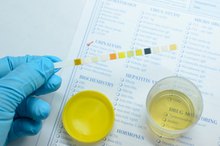
A Diagnosis of an Infection of the Urinary Tract & Lactobacillus
Learn More
Wayson, Wright or Giemsa stain may show up as a safety pin stained shape in the bacterium yersinia pestis, but may also appear as a plump rod or bacilli shape 1. The bacterium stains Gram-negative and cultures can grow at body temperatures of 95 to 98.6 Fahrenheit.
Yersinia pestis is the only species of its genus that remains immotile at room temperature. The bipolar staining is more commonly observed in smears made from clinical specimens rather than cultures.
- Wayson, Wright or Giemsa stain may show up as a safety pin stained shape in the bacterium yersinia pestis, but may also appear as a plump rod or bacilli shape 1.
- The bacterium stains Gram-negative and cultures can grow at body temperatures of 95 to 98.6 Fahrenheit.
Bipolar Staining in Burkholderia Pseudomallei
Burkholderia pseudomallei, a Gram-negative bacterium, is motile, aerobic and rod-shaped like yersinia pestis. Burkholderia pseudomallei can infect both humans and animals to cause melioidosis and glanders.
Under bipolar staining, the bacterium shows up as a Gram-negative intracellular organism. Beyond this, however, the staining characteristic has very little identification value when the bacterium is obtained from clinical samples. Identification must be based on cultures, although this may be prone to error in western countries in which the bacterium is rarely observed.
- Burkholderia pseudomallei, a Gram-negative bacterium, is motile, aerobic and rod-shaped like yersinia pestis.
- Under bipolar staining, the bacterium shows up as a Gram-negative intracellular organism.
Related Articles

List of Social Diseases
Learn More
A Diagnosis of an Infection of the Urinary Tract & Lactobacillus
Learn More

Child With High Fever & Hyperactivity
Learn More

5 Different Types of Bacteria
Learn More
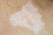
Skin Disorders That Cause Pigment Loss
Learn More
Symptoms of an MRSA Infection on the Face
Learn More

Metronidazole for Acne Treatment
Learn More

What Does Positive Leukocytes Mean?
Learn More

Antibiotics to Treat Cystitis
Learn More

What Are the Causes of Rectal Mucus?
Learn More
References
Writer Bio
Aric Mitchell lives and works out of Fort Smith, Ark. When not writing articles for Demand Studios, he scribes film columns for "Celebrate Arkansas Magazine" and Flickchart.com, and serves as a staff writer for TheRugged.com, a Dallas-based online men's lifestyle magazine.